
Archived news, videos, articles and items of interest.
Return to Current News Items
Follow us on Facebook, Twitter and Instagram.


Is MRI good for examining bones?
We all know both X-rays and CT scans are excellent at visualizing bones, but how do they compare to MRI?
MRI is the best at imaging soft tissues, such as internal organs, brain, muscles, nerves, blood vessels, ligaments and cartilage, but is often overlooked when problems related to the bones are being considered. While both CT and X-Ray are able to demonstrate fractures well, they can both miss small, undisplaced fractures or internal micro-fractures that occur due to trauma or repeated stress. MRI can show these types of fractures before they progress to more serious injuries.
Both CT scans and X-Rays utilize radiation to create their images, while MRI does not use any harmful radiation to acquire images, with the ability to see more types of tissues than either of these modalities. In addition, MRI is often able to characterize these tissues, giving your doctor better information on how to treat these pathologies.
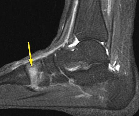
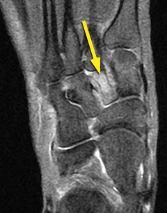
![]()
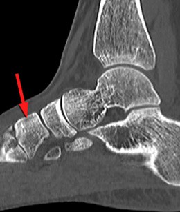
Early stages of a stress fracture of one of the bones of the foot seen on MRI (yellow arrows) but not visible on CT scan (red arrow).
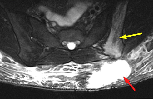
Large subcutaneous contusion (red arrow) with micro-fractures and contusion of the pelvic bone (yellow arrow), not visible on X-rays.
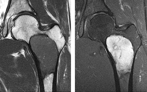
Large hip bone tumor. By using several different types of pulse sequences we can characterize tissues, positively identifying this lesion as benign, which would be treated differently than a malignant tumor.
Please feel free to contact us if you have any questions about how an MRI exam may help your doctor diagnose your health issues. We would be pleased to answer your questions and provide your doctor with information on the MRI exams we can offer.