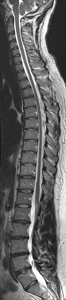
Cervical, thoracic and lumbar spines.
Return to Exam List
Follow us on Facebook, Twitter and Instagram.


| MRI of the Spine | |
|---|---|
| One Area (Cervical/Thoracic/Lumbar/Sacrum) | $975 |
| Two Areas (C-T, T-L, L-S, C and L) | $1,450 |
| Three Areas (C-T-L, T-L-S) | $1,975 |
| Complete Spine (C-T-L-S) | $2,475 |
| C-T-L Spine plus Head Scan | $2,725 |
While many MRI exams of the spine do not require contrast, for those that do, an additional $350 will be charged.
MRI is the preferred method of imaging the spine. Exquisite detail of bones, ligaments, cartilage and the spinal cord with its individual nerve roots are well seen. Pathologies that can be visualized within the spine include fractures, arthritic changes, inflammatory conditions, tumors, MS, traumatic spinal cord injuries, malformations of the blood vessel (AVM or AV Fistula) and disc protrusions.
Your doctor will identify the specific area of your spine from which your symptoms are likely to originate. For example, if you have leg pain and numbness, your doctor might want your Lumbar spine (lower back) to be imaged. Sometimes a larger area that might encompass more areas would be indicated. These include the Cervical spine (neck to upper shoulder blades), Thoracic spine (base of the neck to the bottom of your ribs), Lumbar spine (lower back) or Sacrum (back of your pelvis). Each areas is typically imaged independently, but longer areas may be combined into a "Total Spine" exam. Most single areas of the spine take approximately 30 minutes to image. In some cases you will need the intravenous injection of an MRI contrast agent (dye). We may require you to have a blood test before this procedure. The type of contrast agent we use has been validated to be one of the safest types available. If contrast is necessary, the exam will take an additional 15-20 minutes per section of the spine being imaged.
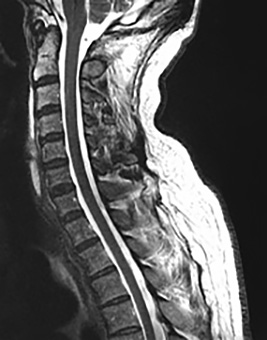
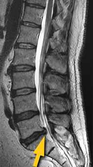
(left) Cervical spine image. (right) Lumbar spine image showing disc protrusion (arrow).
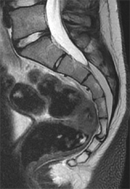
Sacrum and coccyx.