| MRI of the Temporomandibular Joints (TMJ) | $750 |
|---|
MRI is the most accurate non-invasive method of imaging the temporomandibular joints (TMJ). In order to visualize the structure of the TMJs and assess why they may not functioning properly, both sides of the jaw are scanned with the mouth in a closed position, then re-imaged with the mouth in an open position. The entire MRI exam will take approximately 15 minutes.


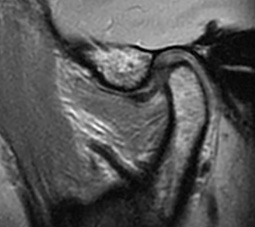
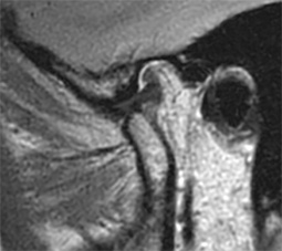
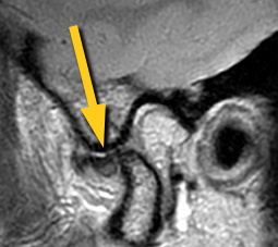
(top left) Normal TMJ in the closed position. (top right) TMJ in the open position, note how the mandibular condyle rides forward with the disc providing padding between it and the articular tubercle. (bottom) Articular disc is anteriorly displaced (arrow), restricting forward motion of the mandibular condyle. This limits the ability to open the mouth.


